By A Mystery Man Writer


All Protocols and Video Articles in JoVE
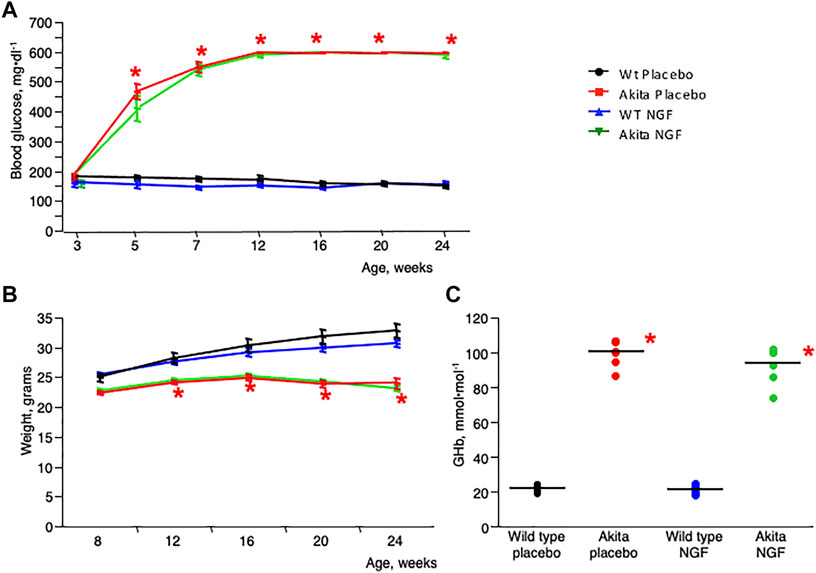
Frontiers Topical nerve growth factor prevents neurodegenerative and vascular stages of diabetic retinopathy
Retina Tool - ImageJ-macros - MRI's Redmine

PDF) Retinal Phenotyping of a Murine Model of Lafora Disease
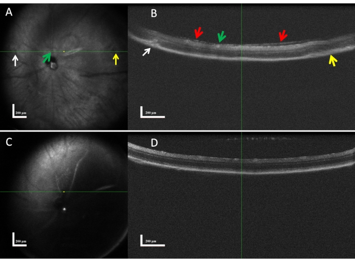
Aplicação da Tomografia de Coerência Óptica a um Modelo de Retinopatia de Rato

Jolanta JAGODZINSKA, PhD Student, Master of Science, Institut des Neurosciences de Montpellier, Montpellier, INM, Vision

All Protocols and Video Articles in JoVE
Frontiers Topical nerve growth factor prevents neurodegenerative and vascular stages of diabetic retinopathy

Topical photodynamic therapy combined with ablative “light needles” against basal cell carcinoma - ScienceDirect

Application of Optical Coherence Tomography to a Mouse Model of Retinopathy

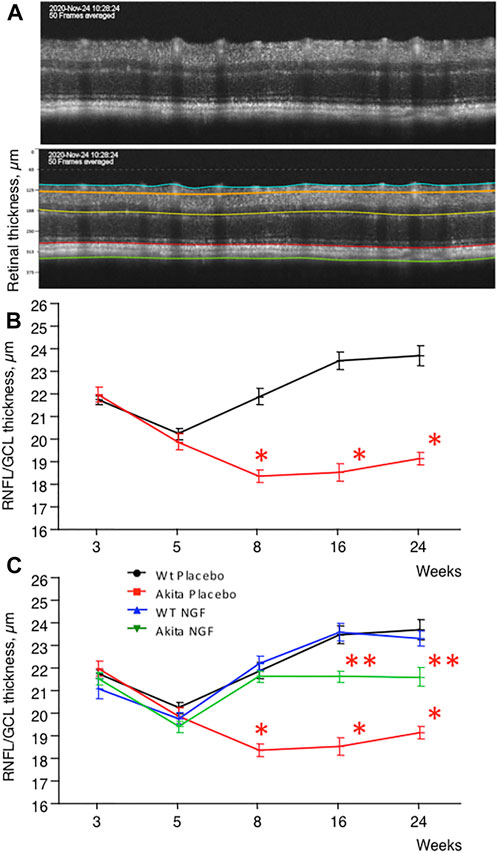
Topical Nerve Growth Factor (NGF) restores electrophysiological alterations in the Ins2Akita mouse model of diabetic retinopathy - ScienceDirect

Topical Nerve Growth Factor (NGF) restores electrophysiological alterations in the Ins2Akita mouse model of diabetic retinopathy - ScienceDirect